
Micro-CT objectives
Non-destructive internal imaging, 3D geometry, pore and void analysis, cracks, inclusions, delamination, and density-related contrast.
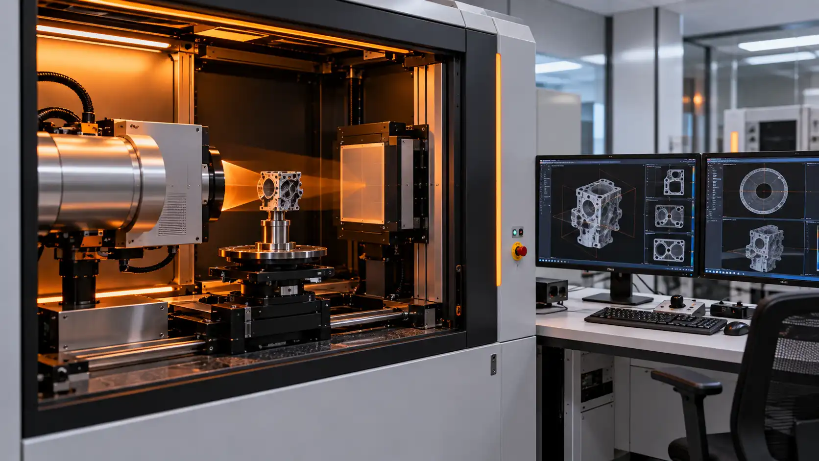
Micro-CT analysis India uses X-ray projections and computational reconstruction to create non-destructive three-dimensional images of internal structures. It can reveal pores, voids, cracks, inclusions, delamination, wall-thickness variation and other density-related features without physically sectioning suitable components, allowing internal geometry and defects to be examined.
Non-destructive internal imaging, 3D geometry, pore and void analysis, cracks, inclusions, delamination, and density-related contrast.
Castings, batteries, electronics, composites, additive-manufactured parts, porous materials, and small engineering components.
Share the sample dimensions, material, expected defect size, density contrast, region of interest, and required 3D output.
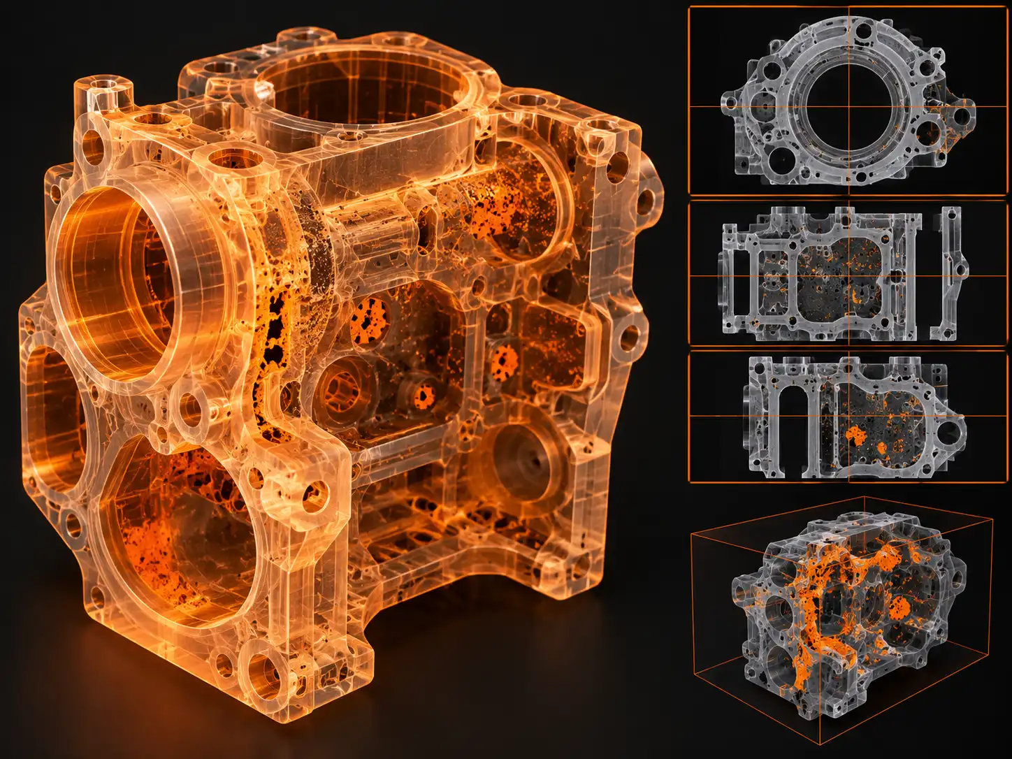
Micro-CT acquires a series of X-ray projections while the sample rotates. Reconstruction algorithms convert the projections into a three-dimensional volume that can be virtually sectioned and inspected.
Results depend on sample size, X-ray attenuation, density contrast, voxel size, scan conditions, reconstruction quality, and the feature scale being investigated.
The appropriate Micro-CT workflow depends on component size, X-ray attenuation, expected defect scale, required resolution, and the internal feature being investigated.
Visualize internal geometry, structures, interfaces, pores, inclusions, and component features in a reconstructed volume.
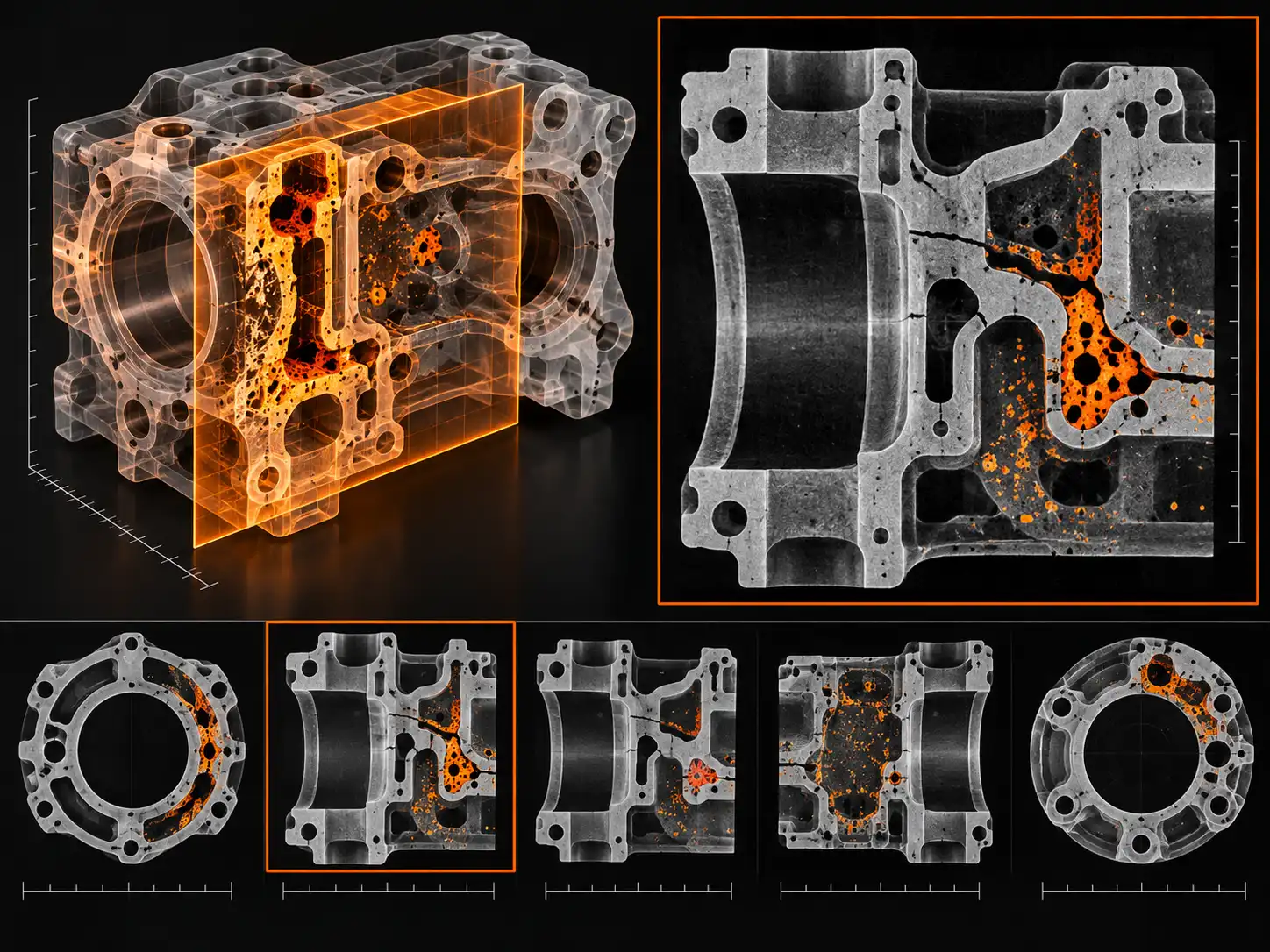
Inspect internal slices without physically cutting the sample to investigate defects, layers, voids, and geometry.
Support porosity, void, crack, wall-thickness, inclusion, dimensional, and internal-structure assessments where contrast permits.
The sample rotates between an X-ray source and detector while a series of projection images is acquired. Differences in X-ray attenuation create contrast between materials, voids, pores, and internal structures.
Reconstruction software combines the projections into a volumetric dataset that can be viewed, virtually sectioned, measured, and analyzed without destroying the component.
Click a workflow component below the diagram to see its role in Micro-CT analysis.
The sample rotates between the X-ray source and detector, projections are acquired from multiple angles, and reconstruction software creates a 3D volume that can be virtually sectioned and measured.
Virtual cross-sections allow an internal plane to be inspected without physically cutting the sample. This can help reveal voids, cracks, inclusions, delamination, internal channels, and geometric deviations.
Interpretation depends on material contrast, voxel size, artefacts, reconstruction quality, segmentation, and the size and orientation of the feature being examined.
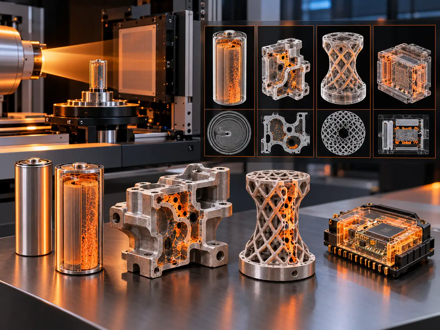
Micro-CT is used when internal structure must be examined without cutting or destroying a component, especially when defects are hidden or three-dimensional geometry matters.
Internal voids, electrode structure, current-collector geometry, cracks, and assembly-related features.
Porosity, shrinkage cavities, inclusions, wall thickness, internal channels, and manufacturing defects.
Internal lattice geometry, lack-of-fusion defects, pores, cracks, and dimensional validation.
Package defects, delamination, inclusions, internal interfaces, fibers, voids, and assembly quality.
Micro-CT performance depends on the relationship between sample size, material attenuation, detector geometry, magnification, voxel size, and the dimensions of the feature being investigated. A smaller voxel does not automatically guarantee that every defect will be resolved or segmented reliably.
Dense materials, strong beam-hardening effects, complex geometries, limited field of view, and low-contrast interfaces can affect reconstruction quality. Scan planning therefore needs to consider the expected defect size, region of interest, material composition, and required measurement accuracy before acquisition.
Micro-CT planning depends on sample size, material attenuation, expected defect scale, density contrast, and the required resolution or output.
Tell us the material, dimensions, internal structure, expected defect, and analytical objective.
Our technical team reviews size, attenuation, density contrast, geometry, and required resolution.
Discuss voxel size, field of view, region of interest, reconstruction, segmentation, and requested outputs.
Receive 3D volumes, virtual slices, measurements, defect observations, and agreed documentation.
Micro-CT is useful when the internal structure matters and the sample provides sufficient X-ray contrast. The final scan route should be selected from the material, dimensions, expected feature size, and desired output.
Inspect internal assembly, pores, voids, electrode geometry, cracks, interfaces, and selected manufacturing features without sectioning the component.
Investigate shrinkage cavities, porosity, inclusions, wall thickness, internal channels, and geometry-related manufacturing features.
Examine lattice structures, lack-of-fusion defects, voids, delamination, fiber-related features, and internal dimensional variation.
Micro-CT analysis is used for non-destructive three-dimensional imaging of internal geometry, pores, voids, cracks, inclusions, delamination, wall thickness, channels, and other density-related features.
Micro-CT may be suitable for castings, batteries, electronics, composites, additive-manufactured parts, porous materials, small engineering components, and other samples with sufficient X-ray contrast and a suitable size.
Micro-CT can support void, pore, crack, inclusion, delamination, and internal-geometry investigations when the feature produces sufficient contrast and is within the effective resolution and field of view.
Micro-CT is generally a non-destructive imaging method because the component can be scanned without sectioning. Suitability depends on the sample, scan conditions, and the required resolution.
Share the sample material, dimensions, expected defect or internal feature, density contrast, region of interest, required resolution, and whether you need 3D reconstruction, virtual slices, dimensional analysis, or defect quantification.
Share a few details for a technical review, or speak directly with our consultant by phone or WhatsApp.
Just a few details — our technical team will guide you.